A eukaryotic-like ubiquitination system in bacterial antiviral defence.
Publication Type:
Journal ArticleSource:
Nature, Volume 631, Issue 8022, p.843-849 (2024)Keywords:
Bacterial Proteins, Bacteriophages, Catalytic Domain, Crystallography, X-Ray, Cysteine, Deubiquitinating Enzymes, Escherichia, Eukaryota, Evolution, Molecular, Lysine, Models, Molecular, Operon, Protein Domains, Ubiquitin-Activating Enzymes, Ubiquitin-Conjugating Enzymes, Ubiquitination, UbiquitinsAbstract:
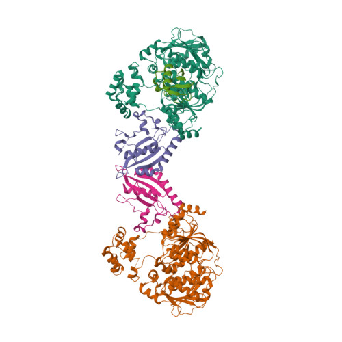
<p>Ubiquitination pathways have crucial roles in protein homeostasis, signalling and innate immunity. In these pathways, an enzymatic cascade of E1, E2 and E3 proteins conjugates ubiquitin or a ubiquitin-like protein (Ubl) to target-protein lysine residues. Bacteria encode ancient relatives of E1 and Ubl proteins involved in sulfur metabolism, but these proteins do not mediate Ubl-target conjugation, leaving open the question of whether bacteria can perform ubiquitination-like protein conjugation. Here we demonstrate that a bacterial operon associated with phage defence islands encodes a complete ubiquitination pathway. Two structures of a bacterial E1-E2-Ubl complex reveal striking architectural parallels with canonical eukaryotic ubiquitination machinery. The bacterial E1 possesses an amino-terminal inactive adenylation domain and a carboxy-terminal active adenylation domain with a mobile α-helical insertion containing the catalytic cysteine (CYS domain). One structure reveals a pre-reaction state with the bacterial Ubl C terminus positioned for adenylation, and a second structure mimics an E1-to-E2 transthioesterification state with the E1 CYS domain adjacent to the bound E2. We show that a deubiquitinase in the same pathway preprocesses the bacterial Ubl, exposing its C-terminal glycine for adenylation. Finally, we show that the bacterial E1 and E2 collaborate to conjugate Ubl to target-protein lysine residues. Together, these data reveal that bacteria possess bona fide ubiquitination systems with strong mechanistic and architectural parallels to canonical eukaryotic ubiquitination pathways, suggesting that these pathways arose first in bacteria.</p>

